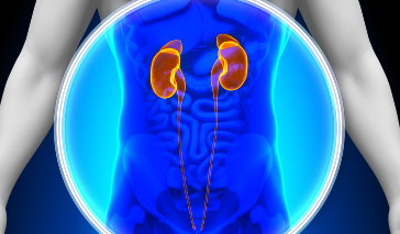
Hydronephrosis occurs when urine collects in the kidneys because it is unable to flow to the bladder. A build-up of urine increases pressure in the kidneys, resulting in distention which can, over time, damage the organs' internal structure. If hydronephrosis develops in one kidney (unilateral hydronephrosis), urinary tract infections or kidney stones may occur. However, if the condition persists in both kidneys (bilateral hydronephrosis), a more serious complication can arise: kidney failure.
Hydronephrosis Causes
Hydronephrosis is not a disease; rather, it is a sign of an underlying condition impacting normal kidney function. As such, it can be caused by an obstruction of the junction between the ureter and renal pelvis (ureteropelvic junction). When this type of blockage occurs, urine is unable to drain from the kidneys to the bladder. Several different conditions can attribute to this, including:
- Congenital abnormalities: Some children are born with structural abnormalities in their renal pelvis or ureteral muscles that cause an obstruction in the ureteropelvic intersection.
- Downward shifting of the kidney (ptosis): Shifting of the kidney due to aging, disease or injury can cause kinking (abnormal bending) at the ureteropelvic interchange, which can hinder the flow of urine to the bladder.
- Kidney stones or blood clots: Kidney stones (calculi) or blood clots in the renal pelvis can block the course of urine from the kidney to the bladder.
- Tumor or abnormal tissue growth: Excess tissue located in the ureteropelvic convergence may compress the ureter, preventing urine flow.
Hydronephrosis can also be triggered by a condition known as reflux. This occurs when urine from the bladder flows back up into the kidneys. Reflux can result from:
- Kidney stones or blood clots in the ureter.
- Tumors or fibrous tissue in or near the ureter.
- Narrowing of the ureter as a result of congenital abnormalities, disease or injury.
- Disorders involving the nerves or muscles of the bladder or ureter.
- Cervix, uterus, prostate or bladder.
- Overactive bladder caused by a spinal cord injury.
Pregnancy can also cause hydronephrosis of one or both kidneys. During pregnancy, the uterus increases in size as the fetus grows. This can compress the ureter, resulting in a blockage of urine flow to the bladder. The condition typically resolves once pregnancy ends. However, treatment for hydronephrosis during pregnancy will be needed to prevent long-term damage to the kidneys.
Hydronephrosis Symptoms
Individuals that develop hydronephrosis may not notice any symptoms until significant damage to the kidney takes place. Additionally, the symptoms that appear will depend on the underlying cause of the disorder and the location of the blockage. For instance, if blockage of both kidneys occurs, the patient may experience renal colic, in which excruciating pain between the ribs and the hip (flank area) occurs. Similarly, patients that suffer from kidney stones may also encounter unbearable pain in the flank area that is intermittent. This pain typically resolves once the stone passes.
Although the specific indicators of hydronephrosis are different for each patient, there are some general symptoms that have been reported. These include:
- Pain in the lower abdomen, back, groin or waist
- Pain when urinating
- Frequent urination typically caused by a urinary tract infection
- Fever/chills
- Nausea and/or vomiting
- Dribbling following urination
- Excessive itchiness of the skin, especially in the groin
Diagnosing Hydronephrosis
In order to diagnose hydronephrosis, a variety of tests must be performed by a doctor. He or she may first conduct a physical exam to identify blockages in the urinary tract before executing a:
- Urine and blood test to determine if kidney damage has occurred
- Abdominal ultrasound to evaluate the structures of the urinary tract
- Intravenous urogram (uses a contrast dye and x-ray to evaluate the function of the urinary tract)
- Cytoscopy (evaluates the lining of the urethra and bladder with a thin viewing instrument known as a cystoscope)
- Voiding cystourethrogram (uses contrast dye and an x-ray to evaluate urinary tract function of the patient while urinating)
Treating Hydronephrosis
Hydronephrosis treatment typically involves resolving the immediate symptoms of the condition and addressing the underlying source of the problem. Draining excess urine from the kidney or treating a urinary tract infection with antibiotics may be the first course of treatment. Once these issues have been attended to, the underlying cause of the problem can be treated. In some instances, such as the development of a kidney stone, no treatment will be needed. In others, such as abnormalities of the ureteropelvic junction, surgery may be required.
References
Hydronephrosis. (2012). Merck Manual. Retrieved from http://www.merckmanuals.com/home/kidney_and_urinary_tract_disorders/obstruction_of_the_urinary_tract/hydronephrosis.html.
LaRusso, L. (2012, October 1). Hydronephrosis—Adult. Conditions & Procedures, 1-3.
Unilateral hydronephrosis (2012). US National Library of Medicine. Retrieved from http://www.ncbi.nlm.nih.gov/pubmedhealth/PMH0001535/.