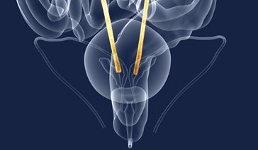
Posterior urethral valve (PUV) refers to an abnormality of the male urethra, the tube in the urinary tract that allows urine to exit the body. During normal fetal development, the urethra and the tissue within it grow, only to stop forming at a certain point. With PUV, the tissue continues to develop beyond the typical growth period, causing a flap to form that restricts the release of urine. As a result, urine flows backwards and builds up within the ureters, bladder, and/or kidneys of the developing fetus, instead of being released into the amniotic sac to assist with lung and other vital organ development. The engorgement of the urinary tract organs can lead to cell and tissue damage, along with a host of symptoms—ranging from mild to severe—that affect urinary function.
Posterior urethral valve treatment options are available, and are aimed at decompressing the urinary tract and restoring urinary function.
Treatment for Posterior Urethral Valve (PUV)
In Utero Treatments
Performing surgery--particularly an endoscopic valve ablation or open surgery--while the fetus is still in the womb is very risky and can lead to complications like limb entrapment, fetal or maternal death, and abdominal injury. Therefore, such an approach is rarely taken.
Rather than take risks, the most common procedures used to treat PUV are minimally invasive and aim to reestablish amniotic fluid, take pressure off of the fetus' urinary tract, and avoid neonatal death. These procedures include:
-
Vesicoamniotic shunt: During this procedure, a cannula on a three-sided needle is inserted through the mother's uterine and abdominal walls, into the amniotic sac and then into the fetus' bladder. A double pig-tailed catheter (usually a Rodeck or Harrison shunt) is then pushed through the hollow center of the cannula and used to connect the distal end of the bladder to the proximal end of the amniotic cavity. The new passageway allows urine to drain into the amniotic sac as it would normally, to assist with lung and organ development.
-
Amniotic fluid infusions: A hollow cannula is placed in the mother's stomach and pushed through until the amniotic sac has been punctured. A tube that looks much like an IV tube, is placed over the opening of the cannula and allows additional amniotic fluids to stream directly into the amniotic membrane.
- Serial bladder aspiration: A needle attached to a syringe is inserted through the mother's abdominal and uterine walls, into the amniotic sac and finally into the bladder of the fetus. The top of the syringe is pulled, which suctions urine out of the bladder.
These fetal interventions are reserved for cases in which severe oligohydramnios (a deficiency of amniotic fluid) is present. A shortage of amniotic fluid can cause the lungs and kidneys of the fetus to be underdeveloped, which can lead to future health problems, such as trouble breathing and renal failure.
Post-Birth Treatments
Following birth, the first step for treating PUV is to relieve pressure on the urinary tract system by inserting a catheter tube into the baby or child's urethra and advancing it up to the bladder. The hollow center of the tube allows urine to drain and exit the body, thus decompressing the bladder and other urinary tract organs. If a urinary tract infection has occurred--a frequent symptom of PUV--antibiotics will then be administered.
Depending on the severity of the condition, surgery may be performed to remove the excess tissue that is causing the blockage and damage to the urinary tract's organs. Surgical procedures for PUV include:
- Valve ablation: Ninety-five percent of PUV cases are treated using this procedure. Here, a specialized tube with a light and camera on its end (cystoscope) is inserted into the urethra. Not only does the device allow a visual of the urethra, but small surgical instruments can be inserted through its hollow center to remove and extract the obstructing tissue.
- Vesicostomy: This is a temporary solution, used when the baby is too tiny for an ablation or the problem is too severe. Here, a hole is made in the bladder, and part of the bladder wall is inverted and attached to the stomach.
- Pyelostomy: An artificial opening in one or both of the kidneys is made so that urine can properly drain.
Prognosis for Posterior Urethral Valve (PUV)
Even after eliminating the problematic tissue for PUV patients, renal failure is not uncommon--up to 35 percent of boys with posterior urethral valve will experience renal failure regardless of the treatment provided. One study, which looked at 70 youngsters over the course of 10 years, found a wide range of problems—again, after the problematic tissue had been removed. These problems included urosepsis, a type of septic poisoning that orginates from an infection in the urinary tract. The study found that the older the child at treatment—older than two years—the worse his related problems were later on.
References:
Urine blockage in newborns. (2010). National Kidney and Urologic Diseases Information Clearinghouse. NIH Publication No. 06-5630
Comprehensive Neonatal Care: An interdisciplinary approach. (2007). Saunders Elsevier.
R K Morris, K S Khan, & M D Kilby. (2007). Vesicoamniotic shunting for fetal lower urinary tract obstruction: An overview. Arch Dis Child Fetal Neonatal Ed.
RA Kukreja, RM Desai, RB Sabnis, et al. (2001). Outcome of children with posterior urethral valves: Prognostic factors. Indian Journal of Urology. Vol. 17.


