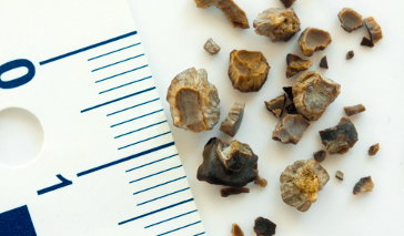
Ureter stones are stones that started their formation in the kidney before moving down to the ureter (a tube that transports urine from the kidneys to the bladder). When materials found in the urine (such as calcium and uric acid) become concentrated, they bind together to form crystals which, over time, accumulate into a solid stone.
Types of Ureter Stones
Stones found in the ureter vary in composition. Identifying their consistency can provide some insight into what is causing them to materialize. Common ureter stones include:
- Calcium stones: Calcium stones are the most common ureter stones and form when there is too much calcium in the urine. High concentrations of calcium in the urine can arise if kidney disease is present. Calcium stones may also form if the intestines absorb excess dietary calcium as a result of intestinal diseases such as Crohn’s disease, ulcerative colitis or inflammatory bowel disease.
- Struvite stones: Struvite stones, which are comprised of magnesium ammonium phosphate, develop when bacterial waste settles in the kidney or ureter, often due to a kidney or urinary tract infection. The ammonia in the bacteria crystallizes and attracts magnesium and phosphate from the urine. This accumulation happens rather quickly, allowing struvite stones to become significant in size.
- Uric acid stones: Uric acid stones are created when the uric acid concentration of the urine becomes elevated. This can happen if there is not enough fluid in the body or if certain health conditions such as gout or rheumatoid arthritis are present.
- Cystine stones: Cystine stones are rare and occur as a result of a genetic condition, known as cystinuria, that causes the kidney to release too much cystine (a crystalline amino acid) into the urine. Much of the cystine that enters the kidneys dissolves and goes back into the bloodstream. However, those with cystinuria have a genetic flaw that interrupts this process. As such, the amino acid collects in the urine and forms crystals or stones that may get lodged in the kidneys, ureters or bladder.
Ureter Stone Risk Factors
Kidney and ureter stones are among the most common and painful urological conditions that develop in adults. If the stone is large enough, it can block the ureter and the flow of urine to the bladder. Doctors do not always know what causes a ureter stone to form, but there are some risk factors that can increase its chance of developing. These include:
- Being male: Men are more likely to develop kidney stones than women because they typically have more muscle mass and eat diets higher in protein. As a consequence, men’s bodies produce more waste products, making them more susceptible to developing ureter stones.
- Family history: Individuals that have a family history of kidney stones are more likely to have this condition.
- Consuming a high-protein diet: Protein is rich in calcium and uric acid. As the concentration of these compounds in the urine rise, so does the threat of developing a ureter stone.
- Being obese: A high body mass index (BMI) is commonly associated with fluctuations in metabolic function. These modifications may lead to higher concentrations of calcium in the urine, which can initiate the formation of calcium stones.
- Intestinal or digestive diseases: Intestinal or digestive diseases can cause changes in the absorption of nutrients and water, leading to the development of ureter stones.
Symptoms of Ureter Stones
Ureter stones can cause a wide range of symptoms that span from mild groin discomfort to excruciating pain. The severity of symptoms will depend on the size of the ureter stone and may include:
- Severe pain that radiates to the side, back, lower abdomen and groin
- Painful urination
- Blood in the urine
- Nausea and vomiting
- Increased urge to urinate
- Increased frequency of urination
- Fever/chills
How Are Ureter Stones Diagnosed?
Not all stones that pass to the ureter will cause symptoms. Some patients are diagnosed after they have had tests (x-ray or ultrasound) performed for other medical reasons. If the ureter stone does cause symptoms, the doctor will conduct a physical exam to determine where the stone is located. In addition, he or she may use the following to confirm diagnosis:
- X-ray of the urinary tract to confirm the location of the stone.
- Ultrasound of the ureter to evaluate the size of the stone.
- Urine tests to determine if an infection is present and to conclude the composition of the stone (calcium, uric acid, etc.).
Treatment for Ureter Stones
Treatment for ureter stones will depend on the size of the stone and whether or not the patient is able to pass it without active treatment. While medications can help alleviate pain during the passage period, shock wave therapy (lithotripsy) may be needed to break the stone apart if it is too large. In more severe cases, such as when there is significant blockage or damage to the renal system, surgery is an option. (Click here to learn more about ureter stone treatment options).
References
Guidelines for the management of ureteral calculi. (2007). European Association of Urology. Retrieved from http://www.klinikverbund-suedwest.de/fileadmin/einrichtungen/KHSIFIBB/Urologie/dokumente/UreteralStonesGL_EurUrol2007.pdf.
Kidney stones and ureteral stones. (2013). Urology Care Foundation. Retrieved from http://www.urologyhealth.org/urology/index.cfm?article=148.
Management of ureteral calculi: Diagnosis and treatment recommendations. (2007). American Urological Association. Retrieved from http://www.auanet.org/content/guidelines-and-quality-care/clinical-guidelines/main-reports/uretcal07/chapter1.pdf.


