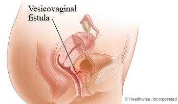
Vesicovaginal fistulas occur when the bladder connects directly to the vagina, leading to loss of bladder control and incontinence.
A fistula is an abnormal passageway between two organs that do not normally connect. Fistulas can appear in most parts of the body and can be caused by trauma, surgery, or can be present at birth.
A vesicovaginal fistula connects a woman's bladder directly to her vagina, which causes urine to drain into the vagina without passing through the urethra and results in loss of bladder control.
Causes of Vesicovaginal Fistula
Vesicovaginal fistulas can be congenital (present at birth) or acquired (developing over time).
Congenital causes are rare and include anomalies that occur during fetal development when the reproductive, intestinal, and urinary tracts merge to create a single pathway.
Acquired causes of vesicovaginal fistulas include:
- Prolonged and obstructed labor during childbirth
- Surgical damage
- Radiation treatment for pelvic cancer
All of the above place stress on the bladder and vaginal wall, which can weaken and tear tissue and lead to a vesicovaginal fistula.
Vesicovaginal Fistula Symptoms
The most prominent symptom is incontinence or urine leakage, with severity depending on the size and location of the fistula. When a fistula is small, some urine still passes through the urethra and may cause only slight leakage. If the fistula is large and all of the urine flows from bladder to vagina, total incontinence can occur.
Even small amounts of urine can irritate the a woman's vulva and perineum if leakage is chronic. When these structures are irritated, a woman may notice pain in her pelvis and lower abdomen and an ammonia-like odor in her urine.
Diagnosis of Vesicovaginal Fistula
Diagnosis is typically made based on a patient's medical history, symptoms, and a physical exam. Urologic tests, such as a urinalysis or urine culture, can confirm diagnosis. Other tests that may be ordered include:
- Intravenous pyelogram: This test uses dye injected into the bladder along with X-rays to visualize the flow of urine through the urinary tract and, potentially, the fistula's location.
- CT scan or MRI: A CT scan uses computer-processed X-rays to generate images, while a MRI utilizes magnetic energy. Both allow a physician to visualize urinary tract organs and determine whether there's an irregular connection.
- Vaginoscopy: During this exam, a tube (cystoscope) equipped with a small camera on the end is inserted into the vagina to view the urinary tract and detect any visible abnormalities.
Treatment for Vesicovaginal Fistula
The size of the vesicovaginal fistula will determine treatment. Fistulas smaller than 3mm in diameter may be closed using estrogen therapy, which strengthens the wall of the vagina. For vesicovaginal fistulas that are larger, surgery may be needed to shut down the pathway. Check out this extended article for more information about vesicovaginal fistula treatment options.
References
Garthwaite, M., & Harris, N. (2010). Vesicovaginal fistulae. Indian Journal of Urology, 26(2), 253-256.
Graham, S.D., Keane, T.E., & Glenn, J.F. (2009). Glenn’s urologic surgery (7th ed.). Philadelphia, PA: Lippincott, Williams & Wilkins.
Slack, A., Jackson, S., & Wein, A. (2008). Fast facts: Bladder disorders. Health Press Limited.