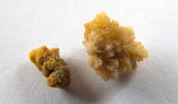
Ureter stones are kidney stones that have been passed to the ureter (a tube that transports urine from the kidneys to the bladder). When certain materials commonly found in the urine (e.g., calcium, uric acid, etc.) become concentrated, crystals form and over time they develop into hard stones that, when lodged in the ureter, can block the flow of urine from the kidneys to the bladder.
The choice of treatment for a ureter stone ranges from watchful waiting to surgery and will depend on the stone's size, as well as the severity of pain it causes.
Watchful Waiting for the Treatment of Ureter Stones
The most conservative treatment option for a ureter stone is watchful waiting, in which no medical treatment to remove the stone is provided for four to six weeks. Rather, the doctor monitors the stone with periodic x-rays or ultrasounds to ensure that it is not growing or transforming. This approach can be effective if the stone size is less than 7 mm in diameter. However, infection and obstruction of the urinary tract are the main difficulties associated with waiting for a kidney stone to make its way out of the body.
Even though watchful waiting is the least invasive method for treating a ureter stone, patients that choose this treatment option may experience tenderness and pain. Typically, the doctor will provide the patient with medications such as ibuprofen, indomethacine, pentazocine or hydromorphone to control discomfort until the stone passes. It is difficult to know for sure if the stone will move through spontaneously. Stones that are less than 5 mm in diameter have a 68 percent passage rate within six weeks. Stones larger than 5 mm in diameter have a 47 percent chance of passing within six weeks.
During watchful waiting, patients will also be advised to consume more water (at least three quarts per day) to increase urine output, pushing the stone to the bladder and out of the body. If signs of an infection develop (e.g., fever, chills, nausea or vomiting), the pain becomes unbearable for the patient or if the ureter stone blocks the flow of urine from the kidneys, watchful waiting may need to be abandoned in favor of more invasive procedures.
Extracorporeal Shock Wave Lithotripsy (ESWL) for the Treatment of Ureter Stones
Extracorporeal shock wave lithotripsy is a minimally invasive procedure that uses shock waves -- applied outside of the body (extracorporeal) -- to shatter the ureter stone into smaller pieces. Once the ureter stone is broken down, the pieces may be more capable of passing through the ureter, bladder and urethra spontaneously. ESWL may cause discomfort for the patient and is often performed with the aid of general or local anesthesia. Although ESWL is highly effective for treating ureter stones, more than one session may be needed to completely remove the stone.
The pass rate for ureter stones when using ESWL has been noted to be as high as 90 percent. However, the location of the stone in the ureter and the size of the stone will impact the success of this procedure. Stones that are larger than 10 mm in diameter and located in the upper region of the ureter have a lower pass rate than those located in the lower ureter with a diameter under 10 mm.
Successful stone passage is also dependent on the health of the patient, how long the stone has been in the body and the composition of the stone. Patients with hydronephrosis (a buildup of urine in the kidney) may be less likely to pass stones even if they are broken up through the use of ESWL. Additionally, the longer stones have been in the ureter, the less likely they are to pass. Stones that have been in the body for long periods of time can become impacted and surrounded by mucosal edema, making it more difficult for them to move through spontaneously, even if they are broken into smaller pieces. Cystine stones are often too hard to shatter with shock waves.
Although ESWL is a highly effective treatment for ureter stones, the procedure does have some potential complications. Stone fragments may block the ureter, causing hydronephrosis or a urinary tract infection. Stone fragments may also remain in the ureter or bladder, growing into larger stones over time. Also, the procedure can cause trauma to the small vessels of the kidney and other structures of the urinary tract. This can lead to renal hematoma or hemorrhage in the short-term and the development of scar tissue over the long-term.
Stones fragmented through the use of ESWL typically pass within a few days. Following the procedure, the patient may experience pain as the stone makes it way through the body. Blood in the urine is also common in the first few days. Studies comparing ESWL and ureteroscopy have not shown notable differences in pass rates or stone-free rates.
Ureteroscopy for the Treatment of Ureter Stones
Ureteroscopy is a more invasive procedure that involves the placement of a ureteroscope into the ureter. The slender viewing instrument is passed through the urethra and the bladder into the ureter to visualize the stone. Once the stone can be seen, the surgeon can insert a flexible basket into the ureter to capture and remove the stone.
Ureteroscopy is frequently used when the stone is located at the lower end of the ureter and is less than 10 mm in diameter, but is too big to pass spontaneously. This procedure can be used in conjunction with ESWL to remove stone fragments that may not be capable of passing through the ureter on their own. Ureteroscopy is an outpatient procedure that typically does not require hospitalization. The patient is usually given general anesthesia and a stent is generally placed in the ureter to ensure that it remains open and heals properly following the procedure.
Although ureteroscopy is a safe and effective procedure for the removal of ureter stones and has a 90 percent clearance rate, some complications may arise. During the procedure, small tears in the ureter may occur. Additionally, detachment (avulsion) of the ureter may result, requiring the surgeon to replace the ureter with an implant. Following the procedure, the patient may experience pain, fever, infection or blood in the urine (hematuria). Stricture or narrowing of the ureter may also take place, demanding the placement a stent to keep the tube open.
Percutaneous Nephrolithotomy (PNL) for the Treatment of Ureter Stones
Percutaneous nephrolithotomy, or PNL, requires the surgeon to make an incision in the flank area of the skin (the area on the back between the ribs and the hip) to access the kidney. A small incision is then made in the kidney so that a thin guide wire can be inserted from the kidney to the ureter. An x-ray is used to visualize the guide wire and ensure that it is properly placed. Once the guide wire reaches the ureter, the surgeon dilates the wire so that a device to remove the stone can be slipped in.
Percutaneous nephrolithotomy is typically used to remove ureter stones that are located in the upper ureter near the kidney and are 1 to 2 cm in diameter. Like ureteroscopy, PNL can also be used in conjunction with ESWL to remove smaller stone fragments that may not be capable of passing through the ureter on their own. It is an invasive procedure that requires general anesthesia for the patient and may result in injury to the colon and blood vessels of the kidney. Following surgery, the patient may experience pain, blood in the urine or infection.
Open Surgery for the Treatment of Ureter Stones
If less invasive efforts to remove the ureter stone are unsuccessful, open surgery may be needed. Open surgery requires a surgical incision in the lower abdomen to access the ureter. During this procedure, the ureter is cut open to directly remove the stone. This is a complex and invasive method that requires at least six weeks for the patient to recover. Following surgery, the patient will be expected to stay in the hospital for five to seven days, at which time they may experience pain and swelling at the incision site. Patients will be advised to increase fluid intake to produce at least two liters of urine per day.
Prognosis for the Treatment of Ureter Stones
Prognosis for the treatment of ureter stones depends on the intervention used. More invasive procedures, like percutaneous nephrolithotomy and open surgery, carry risks such as infection or scarring and may result in additional health problems for the patient. Regardless of the treatment option selected, patients that develop an initial ureter stone are 50 percent more likely to develop another stone within five years.
References
Kidney stones and ureteral stones. (2013). Urology Care Foundation.
Lithotripsy. (2013). US National Library of Medicine.
Ureteroscopy. (2013). National Kidney Foundation.
Surgical management of stones. (2011). Urology Care Foundation.
Preminger G, Hans-Goran T., Assimos D, et al. 2007 guideline for the the management ureteral calculi. European Association of Urology.
Management of ureteral calculi: Diagnosis and treatment recommendations. (2007). American Urological Association.