Contrast radiology is the use of dye, called a contrast medium, which enhances medical images such as X-rays, ultrasound, magnetic resonance imaging (MRI) or computed tomography (CT) scans when certain parts of the body cannot otherwise be easily visualized. The contrast agent may be injected into a vein or directly into the area of the body to be imaged.
Problems that Could Require Imaging of the Urinary Tract
A doctor may order different imaging tests to help find the cause of one or more of the following symptoms:
- Blood in the urine
- Urinary retention—the inability to completely empty the bladder
- Urinary frequency—urinating eight or more times a day
- Urinary urgency—the inability to delay urination
- Urinary incontinence
- Urine blockage
- Urinary tract infection (UTI) with fever
- Repeated UTIs
- Trauma to the bladder
- Swelling in a specific part of the abdomen
- Pain in the groin or lower back
- High blood pressure
- Kidney failure
Each symptom can have several possible causes. In urology, imaging studies can help doctors diagnose urinary tract infections (UTIs), urinary retention, kidney or bladder stones, various kidney diseases, tumors, small bladder capacity, and urinary reflux (the backward flow of urine from the bladder into the kidneys).
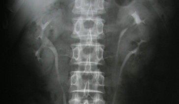
Common Radiology Exams
Computed tomography
CT scans use X-rays sent from numerous angles (which are later processed by a computer or "computed"), often combined with contrast dye, to evaluate the urinary tract and renal system, locate kidney stones, evaluate tumors, plan surgeries, and look for vascular abnormalities.
Intravenous pyelography (IVP)
IVP uses X-rays in combination with an iodine-based contrast medium to assess the kidneys, bladder, and ureters.
Contrast cystography
Using a urinary catheter, a technician fills the bladder with contrast dye. Either still X-rays or fluoroscopy (continuous, moving X-ray images) are taken at various stages of filling and from various angles to visualize the bladder. Contrast cystography is used to check the bladder for rupture after trauma or to look for a vesical fistula (an abnormal passage from the urinary bladder to another part of the body, such as the skin, vagina, uterus, or intestinal tract). Additional images may be taken after drainage of the dye.
Voiding cystourethrography (VCUG)
For this exam, the patient is catheterized, and the bladder and urethra are filled with contrast medium to make the structures clearly visible on the X-ray images. Then, images of the bladder and urethra are taken when the bladder is full and as the patient urinates (voids). This test can show abnormalities inside of the urethra and bladder. VCUG images can indicate how well the bladder empties during urination and whether any urine backs up into the ureters or kidneys (a condition called vesicoureteral reflux).
Retrograde pyelography
This is an X-ray or fluoroscopic technique that is used to evaluate the upper urinary tract, specifically the ureters and kidney. A patient is placed under general anesthesia and a cytoscope is used to visualize the ureters. Contrast dye injected directly into the ureters allows for better visualization. Retrograde pyelography is sometimes used in combination with intravenous pyelography (IVP) to further enhance views of the kidneys and ureters. This imaging technique may be used in patients with blood in the urine (hematuria), or those who have recurrent or suspected urologic cancers (such as renal carcinoma).
Urethrography
Also known as retrograde urethography, this technique is an X-ray examination of the urethra, the tube that carries urine out of the body. A catheter inserted into the urethra is used to inject dye and then X-rays are taken. This is commonly done in men with known (or suspected) trauma or narrowing of the urethra.
Contrast Radiology Complications
Some people can have an allergic reaction to the contrast medium dye and severe allergic reaction requiring medical treatment is rare. Usually, minor reactions include:
- Nausea
- Flushing
- Vomiting
- Itching
References:
ACR Manual on Contract Media. (2012). American College of Radiology Committee on Drugs and Contrast Media. Version 8
Imaging of the Urinary Tract. (2012). National Kidney and Urologic Diseases Information Clearinghouse. NIH Publication No. 12–5107
Indrajit, I.K. (2009). Web review: Contrast Media in radiology and imaging. Indian Journal of Radiological Imaging.
Zagoria, R.J. (2004). Genitourinary radiology: The requisites. Elsevier Health Sciences.