Pyelonephritis is a type of urinary tract infection that affects one (unilateral) or both (bilateral) kidneys. With prompt diagnosis and treatment, patients usually recover without incident. Rarely, pyelonephritis can develop into a serious infection that threatens the afflicted kidney, and possibly the patient’s life. Severe or untreated pyelonephritis may cause permanent kidney scars, which can lead to chronic kidney disease, high blood pressure, and kidney failure.
What Causes Pyelonephritis?
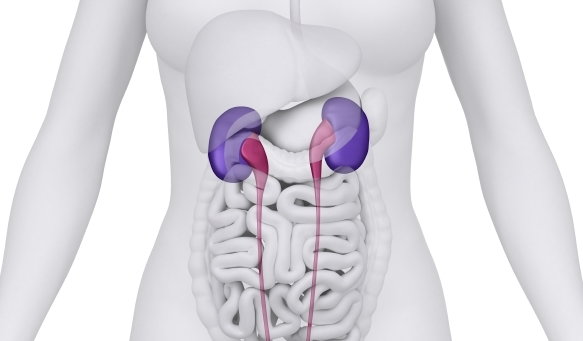
Pyelonephritis results when bacteria or, rarely, viruses infect the kidneys. These bean-shaped organs are located near the middle of the back, just below the rib cage, one on each side of the body. On average, an adult's kidneys filter about 200 quarts of blood every day, producing about 1 to 2 quarts of urine that contain wastes and extra water. Bacteria and viruses can invade the kidneys by moving up from the bladder or by traveling through the bloodstream from other parts of the body. Many bacteria and viruses can cause pyelonephritis, but the most frequent trigger is the bacterium Escherichia coli (E. coli).
Conditions that increase the risk of pyelonephritis include:
- Pregnancy
- An obstruction in the urinary tract (such as a kidney stone or enlarged prostate)
- Urinary system abnormalities (such as ectopic ureters or ureterocele)
- Diabetes
- A compromised immune system
Symptoms Associated with Pyelonephritis?
Symptoms of pyelonephritis can vary and may include the following:
- Frequent, painful urination
- Fever, which can exceed 103°F (39.4°C)
- Chills
- Nausea
- Vomiting
- Back, side, and groin pain
- Blood in the urine, which rarely occurs in males but happens in 30-40% of females with the condition
Children younger than two years old may have no urinary tract-related symptoms and may only exhibit a high fever. In older individuals, pyelonephritis symptoms may manifest as cognitive problems such as confusion, disordered speech, or hallucinations.
How is Pyelonephritis Diagnosed?
Depending on the patient's age, gender and the severity of the symptoms, various tests will be used to diagnose the condition. These tests include:
Urinalysis: During this test, urine is collected in a sterile container and examined at a laboratory. If the urine contains white blood cells and bacteria, there is an infection.
Urine culture: Part of the urine sample is placed in a tube or dish with substances that encourage any bacteria present to multiply. If bacterial colonies grow, something that usually takes one to three days, they can then be identified. Portions of the sample may also be cultured with various antibiotics to determine which drug will most effectively treat the infection.
In most cases, urinalysis is sufficient to diagnose pyelonephritis. However, imaging studies may be required in infants, children, the elderly, and other patients with atypical symptoms. Doctors will also order imaging and other tests if the patient deteriorates or does not respond to therapy within 72 hours. These tests include:
Blood cultures: A blood sample is placed in a tube or dish with substances that encourage any bacteria present to multiply; results typically take one to three days. This test is done to make sure that bacteria have not spread into the bloodstream—a serious condition called sepsis. Blood cultures should be done for any patient being admitted to the hospital for pyelonephritis or who develops the condition when already hospitalized.
Digital (finger) rectal examination: This physical exam of the prostate can determine whether a swollen prostate is obstructing the neck of the bladder.
Ultrasound: Ultrasound uses sound waves to create images of the structure of internal organs. These images can show obstructions in the urinary tract, including physical abnormalities and calculi (kidney or bladder stones).
Computerized tomography (CT) scan: CT scans combine X-rays and computer technology to create 3-D images that are more detailed than those produced by ultrasound. They can show internal organs, as well as obstructions in the urinary tract.
Voiding cystourethrogram (VCUG): A VCUG is an X-ray image of the bladder and urethra taken while the bladder is full and during urination. VCUG can be used to identify abnormalities inside of the urethra and bladder and is usually used to detect vesicoureteral reflux (VUR), a condition in which urine flows backwards from the bladder into one or both ureters and, sometimes, to one or both kidneys, and which can be an underlying cause of recurrent pyelonephritis and other UTIs.
Dimercaptosuccinic acid (DMSA) scintigraphy: DMSA scintigraphy is used to show the severity of kidney infection or kidney damage. During the exam, small amounts of radioactive material are injected into a vein in the patient's arm. A technician uses special cameras and computers to track the substance as it passes through the kidneys. Inflamed or scarred parts of the kidney stand out on the image. DMSA scintigraphy uses less radiation than CT scans.
How is Pyelonephritis Treated?
Pyelonephritis treatment consist of oral antibiotics, typically prescribed for seven to 14 days. Patients who have severe symptoms or an underlying condition are admitted to the hospital and treated with intravenous antibiotics and fluids to prevent dehydration. If a urinary tract obstruction is found, it is often treated with surgery.
References:
Gupta K, Hooton TM, Naber KG, Wullt B, Colgan R, Miller LG, et al. (2011). International clinical practice guidelines for the treatment of acute uncomplicated cystitis and pyelonephritis in women: A 2010 update by the Infectious Diseases Society of America and the European Society for Microbiology and Infectious Diseases. Clinical Infectious Disease.