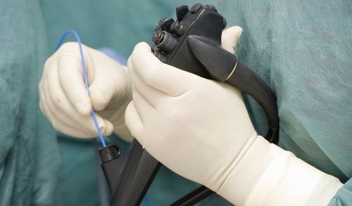
Endoscopy refers to a surgical approach that uses long, thin scopes with a camera and light source at their tip (endoscopes) to see directly inside the body's cavities and organs. The visual the scopes provide can be used to diagnose certain urological diseases and determine the underlying cause behind symptoms. Should a condition (such as kidney stones and bladder stones) present, specialized instruments can be passed through additional channels in the scopes to allow for surgery. In urology, endoscopy is used to examine and treat issues concerning the urinary tract and male reproductive organs.
Types of Endoscopes
Endoscopes can be rigid or flexible. In urology, several types may be used, including:
- Cystoscope - used to examine the bladder
- Ureteroscope - used to examine the ureters, the tubes which allow urine to move from the kidneys to the bladder
- Nephroscope - gives a view of the kidneys internal structures
- Laparoscope - allows a visual of the organs inside the abdomen and pelvis
Endoscopic Approaches
There are two main approaches to urology-based endoscopic surgeries:
Retrograde Endoscopy
This is an approach where the endoscope is placed in the lower portion of the urinary tract and advanced upward. The scope is guided into the urethra (the tube that allows urine to exit the genitals), before being advanced into the bladder and up through the ureters to test for blockages.
If stones, strictures, scar tissue, tumors and other obstructions are found, surgical tools can be guided through an extra channel in the device to cut away and remove them. Repairs can also be made, as can bladder stones be broken up—via laser—in to more extractable pieces. A stent may also be placed in the ureter to help urine move from the kidney to the bladder.
Antegrade Endoscopy
This approach requires a small incision be made in the patient's back so that a scope can be placed in the kidney to look for and gain access to stones, tumors, and other obstructions. Like retrograde endoscopy, the blockages are extracted via specialized instruments that are placed in an additional channel of the scope. If the kidney is not properly draining, a surgeon may leave a tube in the organ to allow urine, stones, and clots to properly drain until function has been restored.
Endoscopy for the Urinary Tract
In urology, a cystoscopy and ureteroscopy are the most common endoscopic procedures performed when diagnosing or treating urinary tract issues.
Cystoscopy
A cystoscope is inserted directly into the urethra (the tube that allows urine to exit the body) and guided up to the bladder. Here, the inner lining of both the urethra and bladder are highlighted, allowing the physician to make a diagnosis. Small surgical instruments—such as laser fibers, graspers, and cutting tools—can be passed through additional channels, making it possible to treat a variety of urinary tract issues. A doctor may perform this procedure if the patient has the following:
- Recurrent urinary tract infections
- Hematuria (blood in the urine)
- Abnormal cells in their urine sample
- An urgent need to urinate
- Chronic pelvic pain or interstitial cystitis/painful bladder syndrome
- Pain during urination
- A urinary tract stone
- A tumor, polyp, growth or cancer in the urinary tract
Ureteroscopy
A ureteroscope is thinner than a cystoscope and is used to reach and visualize the ureters, the two tubes that transport urine from the kidneys to the bladder. The scope can diagnose a variety of urological issues, or channels in the scope allow surgical instruments to be passed through to treat them. This may include:
- Blood in the urine (hematuria)
- An upper urinary tract stone that has moved from a kidney to a ureter
- Unusual narrowing of the ureter (usually causes a blockage)
- Recurrent urinary tract infections
- An abnormal growth, tumor, polyp, or cancer in the ureter
- Odd cells in a urine sample
Endoscopy for the Male Reproductive System
When examining or treating problems with the male reproductive system, the most common procedures performed are:
Trans-urethral Resection of the Prostate:
This procedure treats bladder outlet obstruction due to benign prostatic hyperplasia, which causes the prostate to swell and block urinary flow. A surgeon inserts a rectoscope into the penis and advances it upward to remove excess prostate tissue that is causing the bladder obstruction. After the surgery, a surgeon may recommend continuous bladder irrigation, which involves constantly flushing the bladder with water to remove any excess tissue from the surgery and prevent blood clots from building up in the bladder.
Prostatectomy
When the prostate gland needs to be partially or completely removed due to cancer, it can be done by making several small incisions in the abdomen (laparoscopically). A viewing instrument with a light and camera at its tip (laparoscope) is then inserted into one of the cuts, while surgical tools—including forceps—are guided through other incisions to reach and remove the prostate.
References:
Rastislav H, McNulty M, & Calleary J. (2011). Urology: The home of endoscopy. Prof. Cornel Iancu (Ed.)